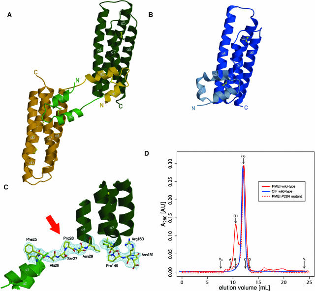
Figure 1.
Structure of PMEI and Comparison with the Invertase Inhibitor CIF.
(A) Ribbon representation of the PMEI dimer with the respective molecules shown in green and yellow.
(B) CIF shown in the same orientation as the green molecule in (A).
(C) The linker region (residues 25PMEI to 29PMEI) interconnecting the dimer as well as a C-terminal extension shown in bonds representation and including the final 2 |Fobs-Fcalc| electron density map (contoured at 1.2 σ).
(D) A 280-nm absorbance trace of an analytical size-exclusion chromatography reveals the presence of PMEI (shown in red) dimers (peak 1) and monomers (peak 2). The invertase inhibitor CIF (shown in blue) appears to be exclusively monomeric. PMEI mutant P28A (dashed red line) does not resemble the dimeric state. Void (V0) and total (Vt) volume are shown for the column together with the elution volumes of molecular weight standards (A, BSA; B, ovalbumin; C, chymotrypsinogen A; D, ribonuclease A). The estimated molecular weight values of the At-PMEI1 monomer and dimer are 19,600 and 37,000, respectively. The calculated monomer molecular weight is 16,400.

