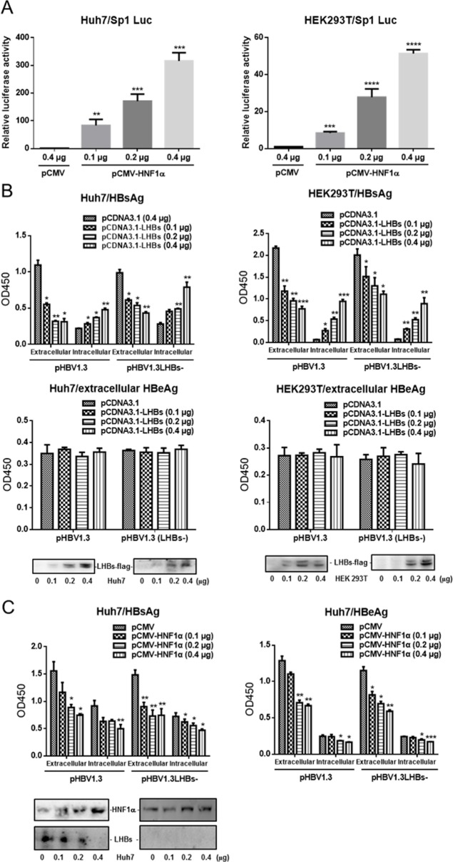
Fig 2. HNF1α’s inhibition of HBV gene expression is independent of LHBs.
(A) Huh7 (left) and HEK293T cells (right) were cultured in 24-well plate and co-transfected with the Sp1 reporter plasmid, pRL-TK and pCMV-HNF1α or pCMV. Means and SEMs of relative luciferase activity data are plotted, with the means of the values from pCMV-transfected cells taken as 1. (B) Huh7 (left) and HEK293T cells (right) were cultured in 24-well plate and co-transfected with the indicated plasmids. The extracellular and intracellular levels of HBsAg and HBeAg were determined. The expression levels of Flag-tagged LHBs were checked with Western blot analysis. (C) Huh7 cells were cultured in 24-well plate and co-transfected with the indicated plasmids. The extracellular and intracellular levels of HBsAg and HBeAg were determined. The virus-derived LHBs was checked with Western blot analysis. Means and SEMs of data from at least three independent tests were plotted. * P <0.05, ** P <0.01, *** P <0.001, **** P <0.0001.

