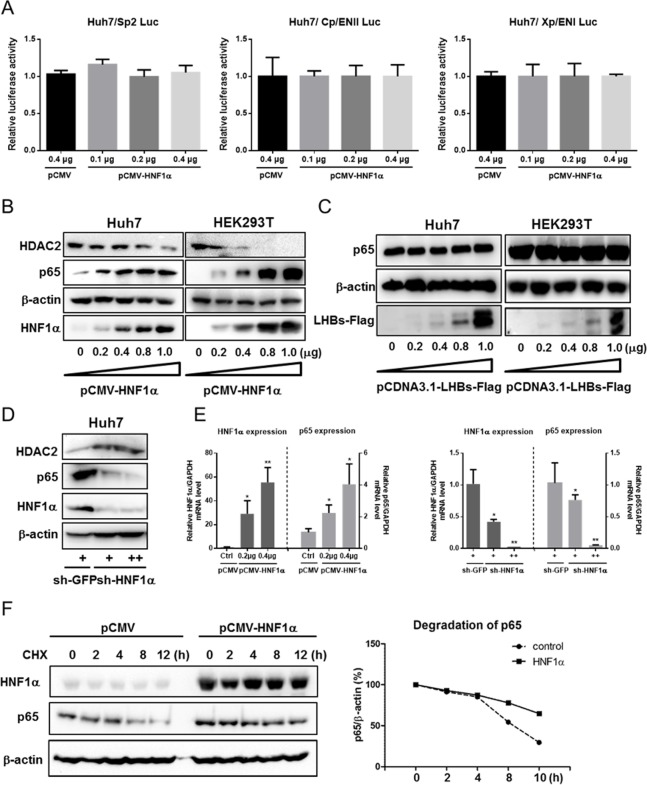
Fig 3. HNF1α promotes p65 expression and protein stability.
(A) Huh7 cells were cultured in 24-well plate and co-transfected with the Sp2, ENII/Cp or ENI/Xp reporter plasmid, pRL-TK and pCMV-HNF1α or pCMV. Means and SEMs of relative luciferase activity data are plotted, with the means of the values from pCMV-transfected cells taken as 1. (B) Western blot analysis of the p65 and HDAC2 levels upon overexpression of HNF1α. Huh7 cells were cultured in 24-well plate and co-transfected with pCMV-HNF1α. (C) Western blot analysis of the p65 level upon overexpression of LHBs. (D) Western blot analysis of p65, and HDAC2 levels in Huh7 cells transduced with the lentivirus expressing sh-HNF1α (+ and ++ stand for 0.2 ml and 0.4 ml lentivirus supernatant per well, respectively) or sh-EGFP. (E) The mRNA expression of HNF1α and p65 were determined with real-time qPCR and normalized against GAPDH. Huh7 cells transfected with pCMV-HNF1α or pCMV (left) or transduced with the lentivirus expressing sh-HNF1α or sh-EGFP. Means and SEMs of data from three independent experiments are plotted with the value of the control takens as 1. *P < 0.05, **P < 0.01. (F) Huh7 cells (1x10⁶) cultured in 12-well plate were transfected with 0.5 μg of pCMV-HNF1α. After treatment with cycloheximide (CHX) for different time intervals, Western blot was performed on cell lysate to detect p65. Protein bands were scanned, quantified and normalized against β-actin. Each time point represents the relative degradation efficiency (%) versus CHX treatment group of 0 hour time point.

