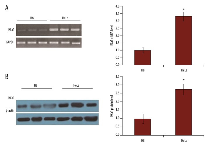
Figure 3.
The expression of IKCa1 mRNA and protein are elevated in HeLa cells. (A) RT-PCR was used to detect IKCa1 mRNA expression with GAPDH as a loading control. (B) IKCa1 protein expression was detected using Western blot in HeLa cells with β-actin used as a loading control. In both A and B (right), mRNA or protein levels were quantified by measurement of the optical density of the samples compared to the optical density of the loading controls. Mean values ±SE of gene or protein expression from 3 independent cell preparations for HeLa and H8. * P<0.05.

