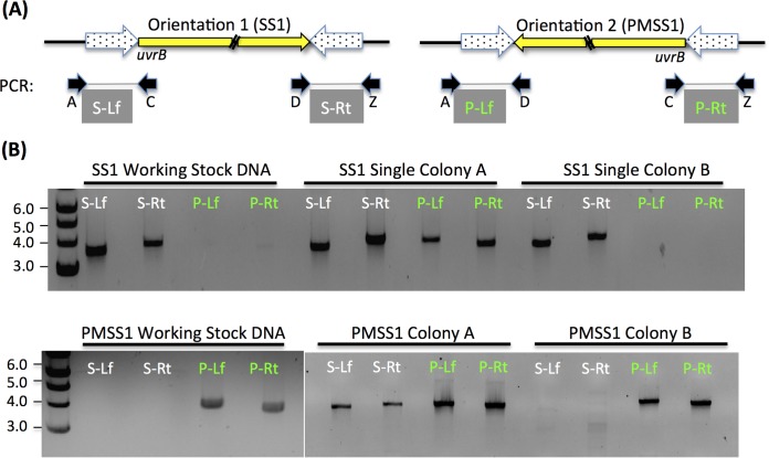
FIG 3 .
A large region variably inverts in SS1 and PMSS1. (A) Diagram showing the orientation of the large inversion region as a yellow arrow. Its size is 425,787 bp. The inversion that was most common in SS1 is shown on the left (Orientation 1), while that in PMSS1 is shown on the right (Orientation 2). The uvrB gene is within the inverted region, just inside one of the inverted repeats (dotted arrows). Primers to confirm the inversion orientation are labeled below each image as A, C, D, and Z. PCR amplification with these yields products A to C (S-Lf) and D to Z (S-Rt) in prientation 1/SS1 and products from A to D (P-Lf) and C to Z (P-Rt) in orientation 2/PMSS1. (B) Gels of PCR amplification products from the primer sets indicated in panel A with template DNA prepared from our working stock H. pylori or from single colonies that were isolated from the freezer stocks and subcultured once prior to DNA extraction. Six single colonies were isolated from each strain, and two representative colonies are shown.

