. 2017 Feb 23;6:e21884. doi: 10.7554/eLife.21884

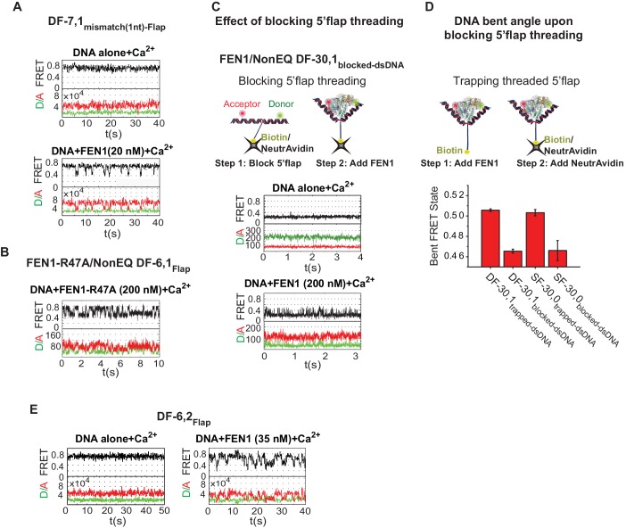
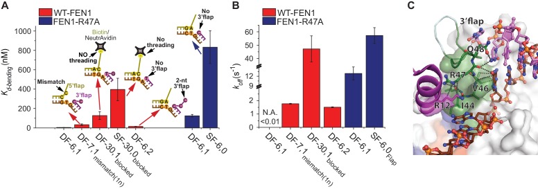
Figure 4. Verification of the bent DNA conformer by the 3’flap-induced protein ordering.

(A) Bar chart comparing Kd-bending for FEN1-WT or FEN1-R47A on various non-equilibrating flap substrates using the internal labeling scheme. Used noncognate substrates include SF-6,0, DF containing 1 nt mismatch at the nick junction (DF-7,1mismatch(1nt)), DF containing biotin-NeutrAvidin on the 5’flap to block 5’flap threading (DF-30,1blocked) and its SF version (SF-30,0blocked), and DF containing 2 nt 3’flap (DF-6,2). (B) Bar chart comparing koff-unbending for FEN1-WT or FEN1-R47A on various non-equilibrating flap substrates using the internal labeling scheme. The lower estimate of koff-unbending for FEN1-WT on DF-6,1 corresponds to the 60 s acquisition time where transitions were rarely detected. Kd-bending and koff-unbending are calculated as in Figure 1—figure supplement 1A and Figure 1G, respectively. koff-unbending was determined from multiple FEN1 concentrations except for FEN1-R47A on SF-6,0 and FEN1 on DF-7,1mismatch(1nt), which were determined from two and one concentration, respectively. The smFRET technique and temporal resolutions used in Figure 4A,B are described in Figure 4—figure supplement 1. (C) R47 acts as a sensor that couples structuring of the 3’flap-binding pocket and the cap-helical gateway. R47 in the hydrophobic wedge mediates multiple interactions, where it stacks against the first base pair on the 3’flap side of the junction while its side chain C-caps the α2 in the gateway (highlighted in green) and stacks with K128 on α5 in the cap (highlighted in purple) (3Q8L.pdb) (Tsutakawa et al., 2011).

DOI: http://dx.doi.org/10.7554/eLife.21884.014

Figure 4.

Figure 4—figure supplement 1. Bending kinetics of various noncognate substrates.

Figure 4—figure supplement 1.

(A) TIRF-smFRET time traces of DF-7,1mismatch(1nt)-Flap alone and in the presence of FEN1. (B) Surface-immobilized confocal-smFRET time traces of NonEQ DF-6,1Flap in the presence of FEN1-R47A at 5 ms temporal resolution. (C) The effect of blocking 5’flap threading on DNA bending by FEN1. Schematic showing the strategy used to block 5’flap threading into the cap-helical gateway by introducing NeutrAvidin/biotin linkage at the 5’end of the 5’flap of DF-30,1 (termed of DF-30,1blocked-dsDNA) prior to the addition of FEN1 (upper panel). Surface-immobilized confocal-smFRET time traces of DF-30,1blocked-dsDNA alone and in the presence of FEN1 at 5 ms temporal resolution (lower panel). The substrate was immobilized by surface-coated-NeutrAvidin via the biotin group on the 5’flap. (D) A bar chart comparing final bent FRET states of DF-30,1trapped-dsNDA, DF-30,1blocked-dsDNA, SF-30,0trapped-dsDNA and SF-30,0blocked-dsDNA using burst confocal-smFRET histograms from freely diffusing substrates acquired at sub-ms temporal resolution. FEN1 concentrations were 5000 nM for SF-30,0internal-blocked, 1000 nM for DF-30,1internal-blocked,1000 nM for SF-30,0internal-trapped and 200 nM for DF-30,1internal-trapped. To trap a threaded 5’flap, FEN1 was first pre-incubated with the substrate before NeutrAvidin was added to bind the biotin on the 5’flap (as shown in the schematic in the upper panel). The FRET value in each condition represents the average of N = 3 and the uncertainty corresponds to the standard error of their fits. (E) TIRF-smFRET time traces of DF6,2Flap alone and in the presence of FEN1.