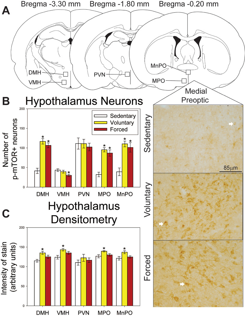
Figure 9.
Effects of exercise on p-mTOR in the hypothalamus. A) Brain atlas diagram modified from Paxinos and Watson (2006) and representative photomicrograms depicting p-mTOR staining in the medial preoptic area (MPO) of sedentary (left), voluntary run (middle) and forced run (right) groups. White arrows indicate examples of p-mTOR-positive neurons. B) Both voluntary and forced exercise increase the number of p-mTOR-positive neurons compared to locked wheel controls in the dorsomedial hypothalamus (DMH), MPO, and median preoptic nucleus (MnPO). Forced wheel running decreases p-mTOR-positive cells in the ventromedial hypothalamus (VMH) compared to sedentary controls. C) Voluntary exercise increases intensity of stain compared to locked wheel controls in the DMH, VMH, MPO and MnPO. Forced exercise does not differ from either condition (p>0.05). * represents p<0.01 relative to sedentary rats. Bars represent group means ± SEM.

