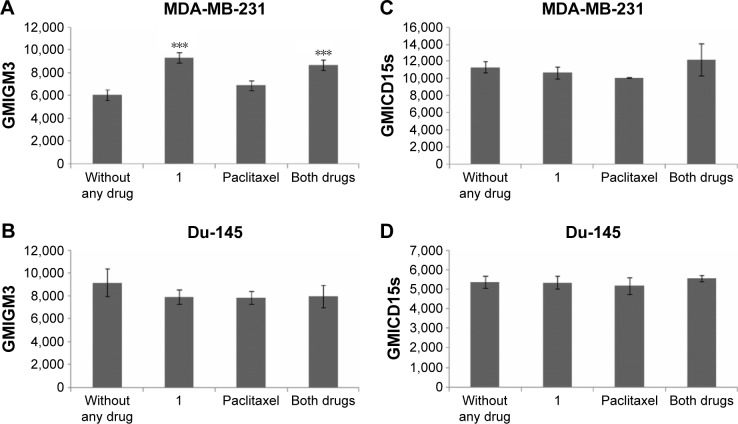
Figure 5.
GM3 and CD15s geometric mean fluorescence of CSC after drug treatment.
Notes: GM3 and CD15s geometric mean fluorescence of CSC after treatment with 2 μM compound 1 combined with 40 or 12 nM paclitaxel, in duration of 48 h in MDA-MB-231 (A, C) or Du-145 (B, D) cell lines, respectively. Data represent the mean ± SD. Columns, mean of viable cells; bars, SD; ***P<0.001.
Abbreviations: CSCs, cancer stem cells; GMI, geometric mean fluorescence intensity; SD, standard deviation.

