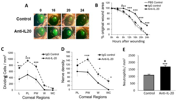
Figure 2.
Corneal wound healing in wildtype mice. (A) Representative images of open wounds revealed by topical fluorescein solution. (B) Percentage of open wound area over time after wounding (n=5, *** p<0.001). (C) Dividing epithelial cells in five regions of the cornea at 24 hours after epithelial abrasion (n=5, ** p<0.01 and *** p<0.001). (D) Nerve density in the epithelium in four regions of the epithelium at 24 hours after abrasion (n=5, * p<0.05 and ** p<0.01). (E) Neutrophil counts in the paralimbal region of the corneal stroma at 24 hours after abrasion (n=5, *** p<0.001). L, limbus; PL, paralimbus; PW, parawound; W, wound margin; WC, wound center.

