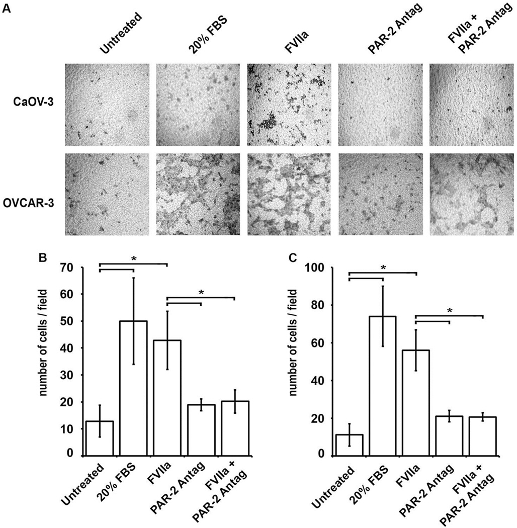
Figure 5. FVIIa induction of EOC migration is PAR-2 dependent.
The effect of pretreatment of serum starved CaOV-3 and OVCAR-3 cells with FVIIa (50 nM) on cellular migration towards 10% serum was analyzed in a trans-well migration assay. (A) Representative images of CaOV-3 and OvCAR-3 cell migration across the insert membrane after 6 hr or 18 hr incubation, respectively, are shown: untreated (negative control), 20% FBS (positive control), 50 nM FVIIa, 1.0 mM PAR-2 antagonist, and 50 nM FVIIa plus 1.0 mM PAR-2 antagonist. Mean migrated cell counts for (B) CaOV-3 and (C) OvCAR-3, corresponding to upper and lower panels in (A), respectively: untreated, 20% FBS, and FVIIa (50 nM) treated cells and inhibition of cell migration after 2 hr pretreatment with 1.0 mM PAR-2 antagonist, and a 6hr or 18 hr incubation, respectively. All results are expressed as mean cell number (n=3) with error bars representing ± SD (*p<0.05).

