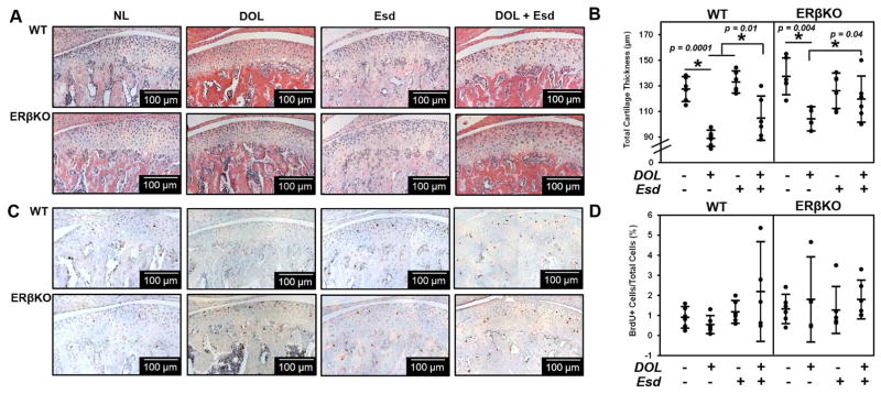
Figure 3. Effect of estradiol and decreased occlusal loading on cartilage thickness and cell proliferation.
The data represent WT and ERβKO mice under normal load (NL) or decreased occlusal loading (DOL) with either placebo (Plb) or estradiol (Esd) treatment. Specifically, the labels indicate the following: NL = normal load and placebo; DOL = decreased occlusal loading and placebo; Esd = normal load and estradiol; DOL + Esd = decreased occlusal loading and estradiol. Representative hematoxylin & eosin (H&E) images (A), cartilage thickness as determined by histomorphometry (B), BrdU proliferative immunohistochemical staining (C), and quantified percentage of BrdU+ cells (D) are shown. For histomorphometric and BrdU analysis, n=6 mice were utilized for all groups and the average of 3–6 sections/mouse was analyzed. BrdU+ cells were normalized to the total number of cells in the cartilage region as determined from the adjacent H&E section. Statistical significance was determined by a two-way ANOVA followed by posthoc analysis with the Bonferonni method with p < 0.05. Exact p values are listed above the bars that denote significance.

