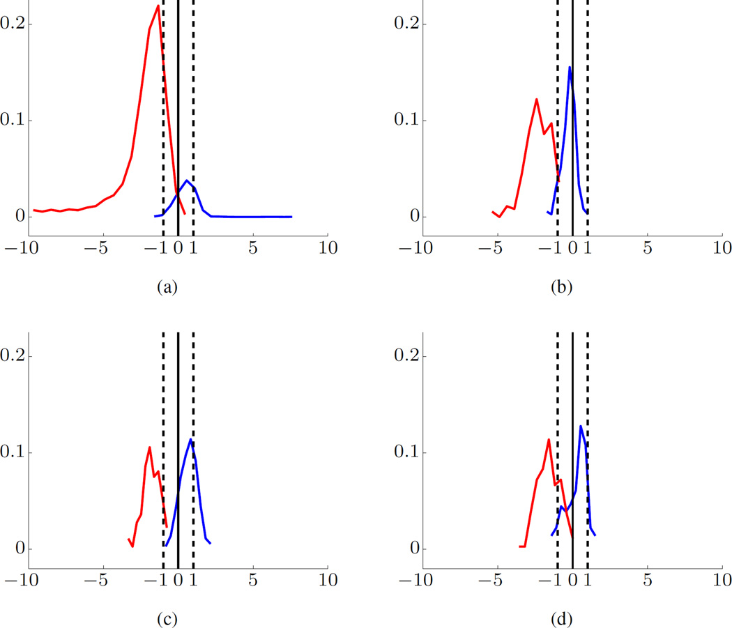
Fig. 4.
SVM modeling for Dog-L4 data set using BFB feature encoding. Histograms of Projections for training data (a), and for test data (b)–(d). The preictal data are shown in blue and interictal data in red. Margin borders correspond to −1/+1 (marked by dashed vertical lines). The x-axis is the scaled distance and y-axis the fraction of samples.

