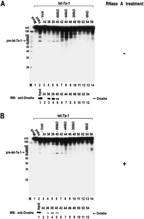
Figure 3.
Gel exclusion chromatography of Drosha. (A) Nuclear extract was prepared from twenty-five 10-cm plates of HEK293T cells and fractionated through a Sephacryl-S300 HR column. (Top panel) Each fraction was 1.75 mL in volume, from which 20 μL was taken for in vitro processing of pri-let-7a-1. The fraction number and the protein molecular mass standards (Sigma) are indicated at the top of the gel. “Mock” means that lysis buffer was used instead of protein fractions for the in vitro processing assay. (Bottom panel) The fractions were also analyzed by SDS-PAGE, and Drosha proteins were detected by Western blotting using anti-Drosha antibody. (B) Nuclear extract was treated with 50 μg/mL of RNase A for 30 min at 4°C before loading to the column.

