Figure 3.

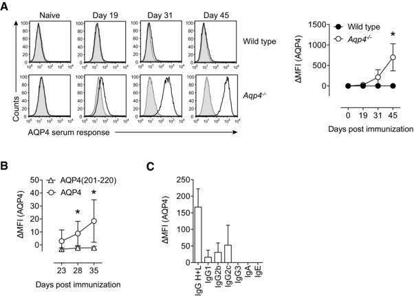
Aqp4 −/− mice but not WT controls mount a robust antibody response to AQP4 upon immunization with full‐length AQP4 protein. WT C57BL/6 and Aqp4 −/− mice were immunized s.c. with full‐length AQP4 protein or AQP4(201–220) peptide emulsified in CFA. (A) Sera of naive mice and AQP4‐immunized WT or Aqp4 −/− mice at different time points after immunization with AQP4 were analyzed for AQP4‐specific antibodies in a cell‐based flow cytometric assay with LN18 cells transduced with AQP4 expressing lentivirus. Anti‐mouse total IgG H+L (AlexaFluor488 labeled) was used to detect anti‐AQP4 antibodies (black line histograms). The ΔMFI was calculated in relation to staining of LN18 cells transduced with empty vector (shaded histograms). Representative histograms (left) and plot of ΔMFI (right). Mean ΔMFI ± SD (n = 6 per group). (B) AQP4 serum response at different time points in Aqp4 −/− mice that were immunized with AQP4 protein or AQP4 (201–220) peptide. Mean ΔMFI ± SD (n = 6 per group). (C) To specify the antibody classes and subclasses, fluorochrome‐labeled anti‐mouse Ig antibodies specific for IgA, IgE, IgG1, IgG2b, IgG2c, and IgG3 were used. Mean ΔMFI ± SD (n = 6). *p < 0.05 (unpaired Student's t‐test). Data are representative of three independent experiments (A–C).
