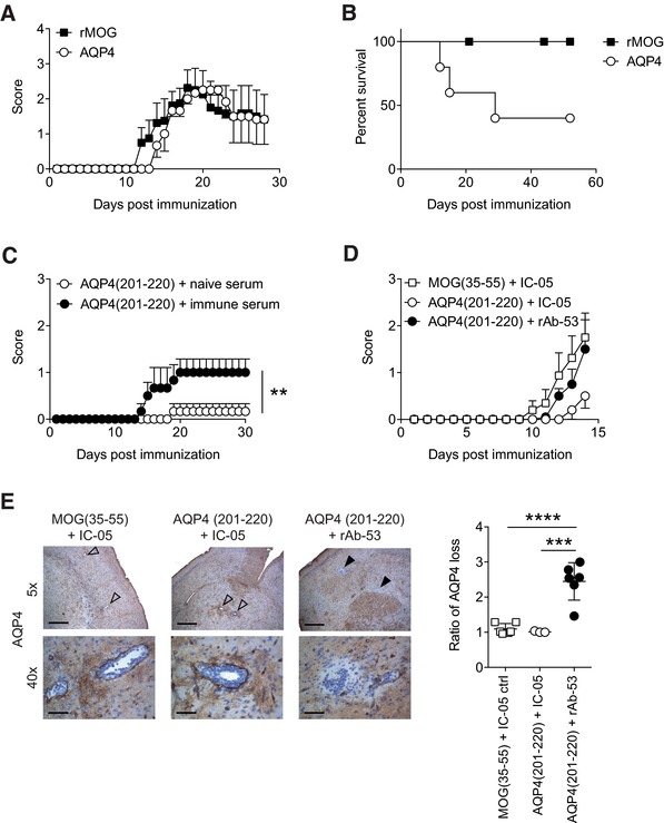
Figure 5.

AQP4(201–220) is an encephalitogenic epitope. (A, B) Aqp4 ΔT × Tcra −/– mice were immunized with full‐length AQP4 or rMOG protein emulsified in CFA and monitored for symptoms of encephalomyelitis. (A) Mean clinical scores ± SEM and (B) percent survival of immunized mice are shown (n = 6 per group). (C) Aqp4 ΔT × Rag1 −/− mice were immunized with AQP4(201–220) peptide emulsified in CFA. One hundred microliters of naïve serum or immune serum with high titers of anti‐AQP4 antibodies (harvested from AQP4‐immunized Aqp4 −/− mice) were administered i.v. on days 6 and 12 after immunization. Mean clinical scores ± SEM (n = 5 per group). **p < 0.01 (Mann–Whitney U test for nonparametric values). (D, E) Aqp4 ΔT × Rag1 −/− mice were immunized with either AQP4(201–220) or MOG(35–55) peptide emulsified in CFA. On days 12 and 14 after immunization, the mice were injected i.v. with rAb‐53 antibody or IC‐05 control antibody. (D) Mean clinical scores ± SEM (n = 6 per group). (E) Mice were sacrificed 4 h after the last antibody treatment to perform histological analysis. Representative AQP4 staining of the brain at 5× (scale bar, 400 μm) and 40× magnification (scale bar, 50 μm). Open arrows show vessels without perivascular loss of AQP4 immunoreactivity. Closed arrows indicate areas of AQP4 loss in the vicinity of vessels. Quantification of AQP4 loss as ratio of the area with AQP4 signal loss divided by the area of the associated vessel lumen in the brain of the indicated treatment groups (mean ± SD). ***p < 0.001; ****p < 0.0001 (ANOVA plus Sidak's post‐test). Data are representative of two independent experiments (A, B, D, and E).
