Fig. 17.

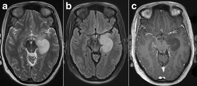
Low-grade glioma. 1.5-T axial FLAIR (a), T2W (b) and contrast-enhanced T1W images (c) show a T2-hyperintense, T1-hypointense, non-contrast-enhancing infiltrative mass in the left hippocampus, corresponding to a pathologically proven glioma

Low-grade glioma. 1.5-T axial FLAIR (a), T2W (b) and contrast-enhanced T1W images (c) show a T2-hyperintense, T1-hypointense, non-contrast-enhancing infiltrative mass in the left hippocampus, corresponding to a pathologically proven glioma