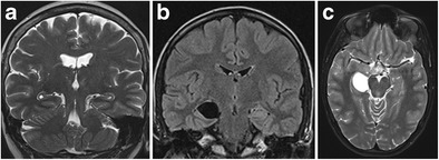
Fig. 6.

Bilateral sulcal remnant cysts (a) and right-sided choroid fissure cyst (b, c). Coronal T2 shows small bilateral cysts at the apex of the hippocampal fold between the dentate gyrus and Ammon’s horn (a). Coronal FLAIR (b) and axial T2-weighted (c) images show a space-occupying cystic lesion, iso-intense to cerebrospinal fluid, at the level of the right choroid fissure
