Fig. 3.
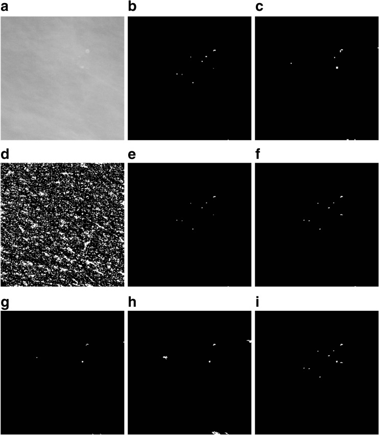
An illustration of detecting microcalcifications in an image 512 × 512 pixels in size, extracted from mammogram A_1131_1.RIGHT_MLO. a The result of microcalcifications detection in stage 1. b, c The result of using the detector based on Eq. (1) at the second and third levels of the morphological pyramid, respectively. d Extended maximum emax. e An image showing the marker for reconstructing microcalcifications detected at the second level of the morphological pyramid—an intersection of images from items (b) and (d). f The result of the reconstruction by dilation of the mask presented in item (d) and the marker presented in item (e), i.e., an image presenting microcalcifications detected at the second level of the morphological pyramid. g An image showing the marker for reconstructing microcalcifications detected at the third level of the morphological pyramid—an intersection of the images from items (c) and (d). h The result of the reconstruction by dilation of the mask presented in item (d) and the marker presented in item (g), i.e., an image presenting microcalcifications detected at the third level of the morphological pyramid. i The sum of images from items (f) and (h), using the OR operator, as the result of extracting microcalcifications using the morphological pyramid. The image has been subjected to additional “cleaning” operations

