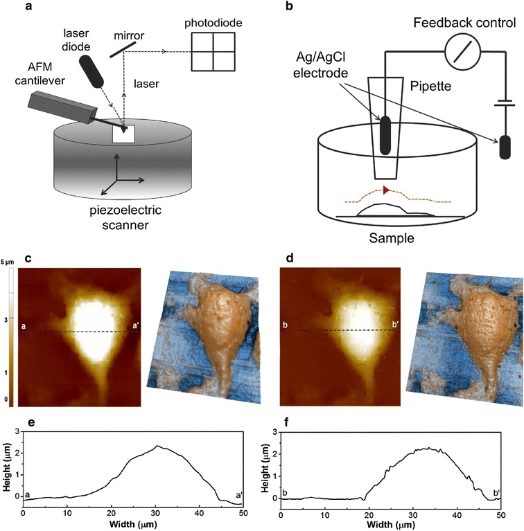
Fig. 1.

Schematic view of principle of SPM techiques and L929 cell surface images using SICM. a In AFM, the attractive or repulsive force between the tip and the sample causes deflection of the cantilever. As the cantilever deflects, the angle of the reflected laser beam changes angle and strikes a different part of the photodiode. The signals from the four quadrants of the detector are compared to calculate the deflection signal. Using this signal, the system (computer) generates a topography of the sample surface. b In SICM, a nano-pipette filled with electrolyte is brought in proximity to the sample of interest. A bias applied between an electrode in the pipette and another electrode in the bulk solution generates an ion current, which can be used in feedback control to prevent direct contact between the nano-pipette and the sample. c Height image and 3-dimensional image of live single L929 fibroblast cell surface using SICM hopping mode, d height image and 3-dimensional image of fixed fibroblast cell surface. The size of all images are 50 × 50 µm, after imaging of live cell (c), fixed with PFA, fixed cell imaging (d) was performed. e, f Indicates line profile of each image. Imaging time of each image is around 30 min
