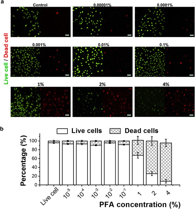
Fig. 6.

Evaluation of PFA-mediated cytotoxicity on L929 cells. a Fluorescence microscope images for assessment of live and dead cell ratio dependent on titration of C PFA. Green fluorescence represents live cells and red fluorescence represents dead cell, scale bar 50 μm. b Percentage graph of live/dead cell ratio dependent on the titration of C PFA. Data is presented as the mean ± standard deviation, with t test results indicating p < 0.05
