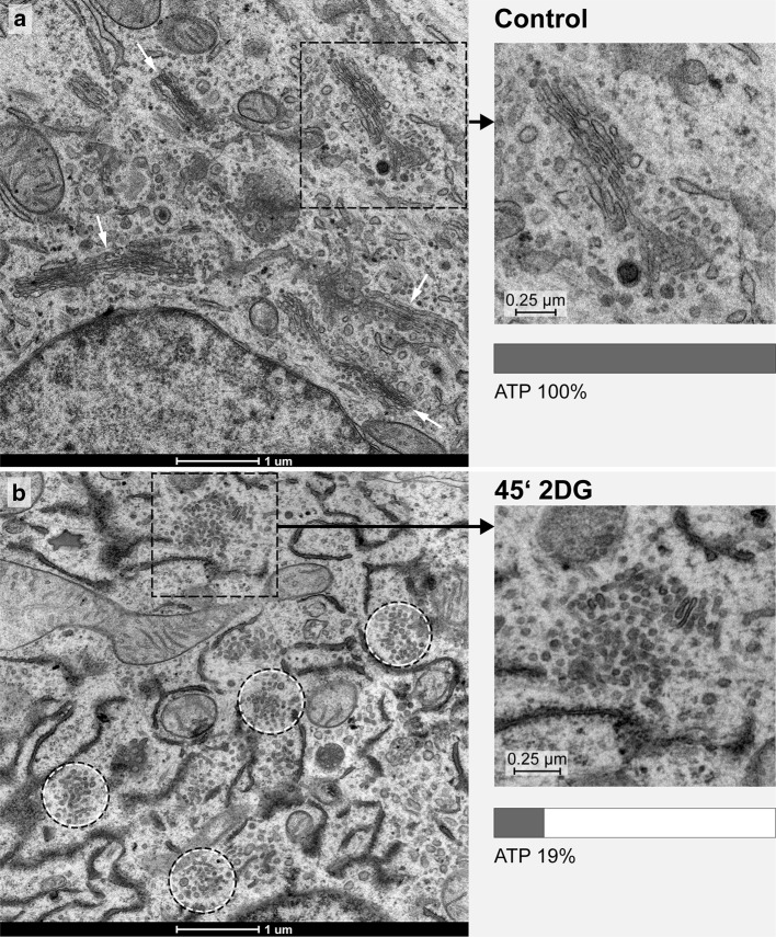
Fig. 1.
Ultrastructures of the Golgi apparatus in HepG2 hepatoma cells, high-pressure frozen, freeze substituted and embedded in Epon, are shown on thin sections of a control cell (a, inset) and after 45 min of treatment with 2DG (b, inset). In the control cell (a), several regular stacks of cisternae in parallel organization are apparent (white arrows); in the 2DG-treated cell (b), regular Golgi stacks are missing and instead Golgi bodies composed of irregular and loosely organized membranous compartments dominate (circles). In both panels, areas marked by a rectangle are shown in the inset at higher magnification and respective ATP-values are indicated. In all pictures, RER-cisternae are found close to the Golgi stacks and Golgi bodies; the RER luminal contents appear denser in the 2DG-treated cells (b) than in the controls (a)

