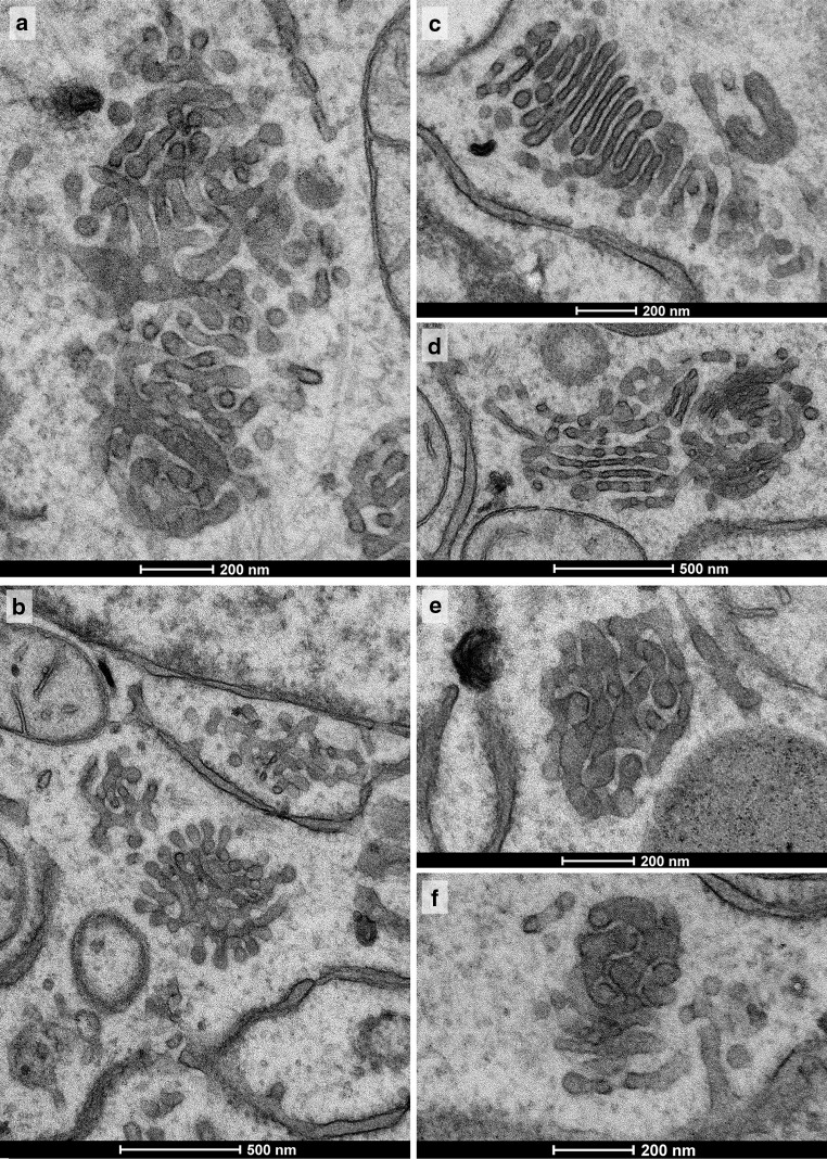
Fig. 10.
All panels show Golgi apparatus bodies after 2DG-removal following a 45 min treatment time and a 30 min subsequent incubation in GPF. Various types of Golgi bodies are on display, as they also may reside in cells side by side. The bodies shown in a, b are of tubular-reticular character; e, f particularly compact glomerular Golgi bodies with densely packed membranes, and c and d initial stack formations. c Multi-cisternal mini-stack, as is characteristic for this recovery period. The cisternae are conspicuously short, lack pores and are in a ladder-like arrangement. A combined body is shown in d consisting of a compact part with densely packed membranes on the right-hand side; in the stack on the left-hand side, pores can be seen and pairs of cisternae appear connected at their rims. Narrow, particularly regular inter-membrane spaces are apparent in the compact body shown in f

