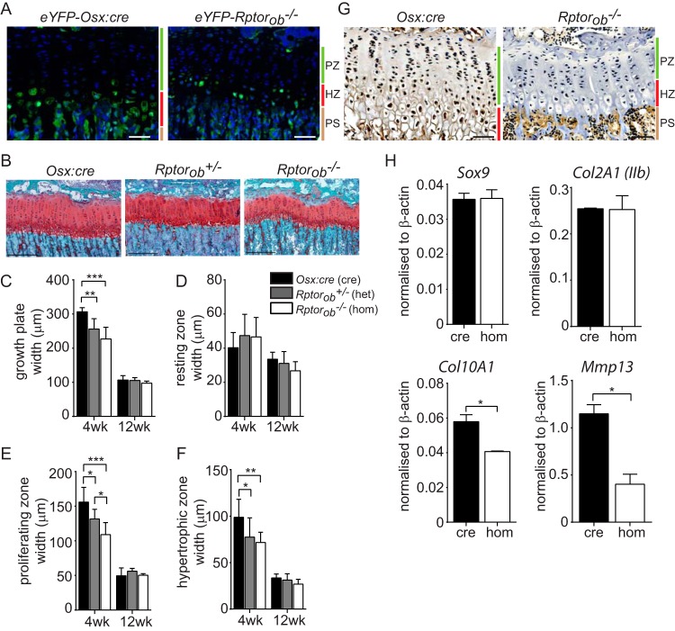
FIG 6.
Narrow and disorganized growth plate in Rptorob−/− mice. (A) eYFP-labeled cells within the hypertrophic chondrocyte zones of epiphyseal growth plates of 4-week-old eYFP-Osx:cre and eYFP-Rptorob−/− mice. Nuclei were stained with DAPI. Scale bar, 50 μm. (B) Safranin O staining of acidic proteoglycan in the epiphyseal growth plate of the tibia. Scale bar, 200 μm. (C) Growth plate widths based on Safranin O staining. (D to F) Width of chondrocyte zones within the growth plate based on chondrocyte morphology. (G) Immunohistochemical staining of PCNA in the growth plates of Osx:cre and Rptorob−/− mice. Scale bar, 50 μm. (H) Quantitative PCR was performed on RNA extracted from eYFP+ cells isolated from eYFP-Osx:cre and eYFP-Rptorob−/− mice using primers specific to the indicated genes. The relative transcript levels were normalized to β-actin. PZ, proliferating zone; HZ, hypertrophic zone; PS, primary spongiosa. Measurements are presented as means ± the SD (n ≥ 6 per group). *, P < 0.05; **, P < 0.01; ***, P < 0.001.

