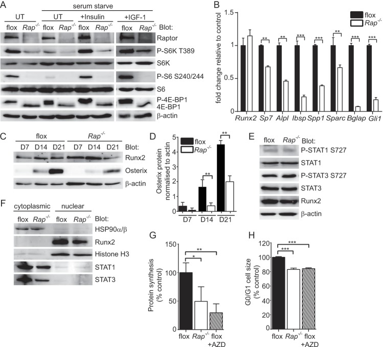
FIG 9.
Mechanisms leading to low bone mass in Rptorob−/− mice. Calvarial cells isolated from Rptorfl/fl mice (P4) were infected with a lentivirus harboring a tamoxifen-inducible cre recombinase. Infected cells were treated with tamoxifen (Rap−/−) to stimulate Rptor gene deletion. Control cells (flox) were treated with vehicle. (A) The effect of Rptor gene deletion on mTORC1 substrate activation at steady-state and following stimulation with either insulin or IGF-1 was assessed by Western blotting. (B) Assessment of osteogenic gene expression in vehicle (flox)- or tamoxifen (Rap−/−)-treated cells after 21 days of osteogenic induction. (C) Western blots showing Runx2 and osterix protein levels in vehicle (flox)- or tamoxifen (Rap−/−)-treated cells after 7, 14, and 21 days of osteogenic induction. (D) Densitometry showing osterix protein levels normalized to β-actin in vehicle (flox)- or tamoxifen (Rap−/−)-treated cells after 7, 14, and 21 days of osteogenic induction. (E) Western blots showing the activation status of STAT1 and STAT3 in vehicle (flox)- or tamoxifen (Rap−/−)-treated cells. (F) Western blots showing the subcellular localization of the indicated proteins in vehicle (flox)- or tamoxifen (Rap−/−)-treated cells. (G) Metabolic labeling in vehicle (flox), tamoxifen (Rap−/−)- or vehicle plus AZD8055 (flox+AZD)-treated cells. (H) Cell size analysis in vehicle (flox)-, tamoxifen (Rap−/−)-, or vehicle plus AZD8055 (flox+AZD)-treated cells. Measurements are presented as means ± the SD from three independent experiments. *, P < 0.05; **, P < 0.01; ***, P < 0.001.

