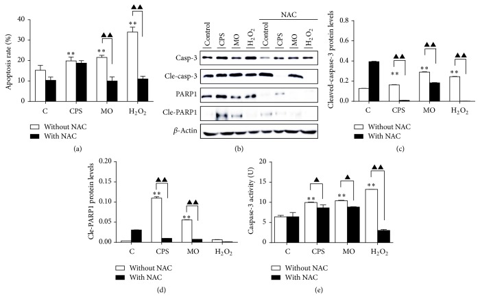
Figure 3.
CPS-induced apoptosis in ALI cultures of sheep bronchial epithelial cells. Cells were pretreated with/without NAC (10 mM) for 2 h, followed by exposure to indicated conditions for 48 h. (a) 4-week-old ALI cultures of sheep bronchial epithelial cells were apically infected with CPS at 100 ng/ml and MO at MOI of 30 for 48 h before samples were harvested for analysis. Percentages of apoptotic cells were determined by flow cytometry using Annexin V/PI double-staining assay. (b) Immunoblots of apoptosis-associated proteins. The blots were probed for β-actin as a loading control. (c and d) Representative blots for cleaved-caspase-3 and cleaved-PARP1 were semiquantified by a densitometric analysis by calculating the fold of change of a protein of interest over β-actin. (e) Relative caspase-3 activity of ALI cells was detected after treating with indicated conditions. Values are mean ± SD for at least three independent experiments performed in triplicate. Compared to non-CPS or MO treatment, ∗p < 0.05 and ∗∗p < 01. Comparing between indicated groups, ▲p < 0.05 and ▲▲p < 0.01.

