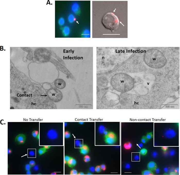
FIG 1.

Horizontal transfer of Wolbachia bacteria between Drosophila cells. (A) Wolbachia bacteria extracted from infected JW18 cells were added to JW18-DOX cells and incubated for 24 h. (B) Wolbachia bacteria extracted from infected LDW1 cells were added to LDW1-DOX cells and incubated for 1 h. (C) Uninfected Drosophila S2 cells and Wolbachia-infected JW18 cells were cocultured on a glass coverslip for 24 h. Wolbachia infections in previously uninfected cells can be seen with FISH (A) and DIC (C) imaging or electron microscopy (B) to determine if horizontal transfer of infection took place. Results are typical of the multiple fields of view examined. Red, Wolbachia; blue, nuclei stained with DAPI; green, GFP-Jupiter (JW18 only). hc, host cell; n, nucleus; v, vesicle; w, Wolbachia. Bar, 10 μm.
