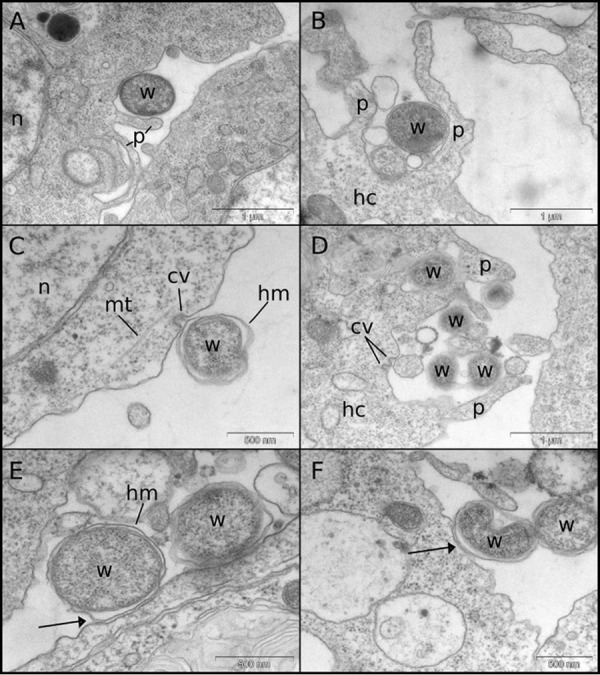
FIG 5.

Transmission electron micrographs of LDW/JW18 cells exposed to Wolbachia bacteria from cell lysates or infected JW18 cells. (A and B) Wolbachia bacteria are frequently seen surrounded by phagocytic pseudopodium-like extensions of the host cell. (C and D) Wolbachia bacteria can be seen contacting what appear to be clathrin-coated pits, sometimes coinciding with pseudopodia (D). (E and F) The host-derived membrane surrounding the Wolbachia double membrane can be seen in close contact with the host cell membrane (arrows). cv, clathrin vesicle; hc, host cell; hm, host membrane; mt, microtubules; n, nucleus; p, pseudopodia; w, Wolbachia.
