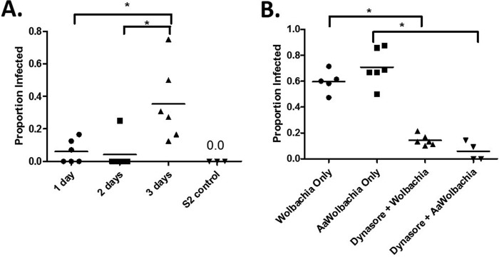
FIG 6.
Horizontal transfer of Wolbachia bacteria takes places between mosquito and Drosophila cells. (A) Uninfected Drosophila S2 cells were seeded beneath Wolbachia-infected A. albopictus cells (C6/36) in a transwell insert. After being cocultured for 1, 2, or 3 days, new Wolbachia infections in previously uninfected cells were visualized by FISH in 6 fields of view for each group. S2 cells plated in the absence of C6/36 cells served as a control for FISH staining. Data are presented as proportion of infected cells ± SEM and were analyzed by one-way ANOVA, followed by Newman-Keuls multiple-comparison test to determine differences between time points (F = 7.78, R2 = 0.509, df = 17). Values were deemed significant when P < 0.05 (indicated by an asterisk above the bracket). (B) Pretreatment of JW18-DOX cells with dynamin prior to the addition of crude Wolbachia preparations from infected Drosophila JW18 cells or mosquito C6/36 cells (AaWolbachia) for 24 h resulted in a reduced ability of Wolbachia bacteria to invade cells. Data are presented as proportion of infected cells ± SEM and were analyzed by one-way ANOVA, followed by Newman-Keuls multiple-comparison test to determine differences between groups (F = 61.4, R2 = 0.912, df = 20). Values were deemed significant when the P value was <0.05 (indicated an asterisk above the bracket).

