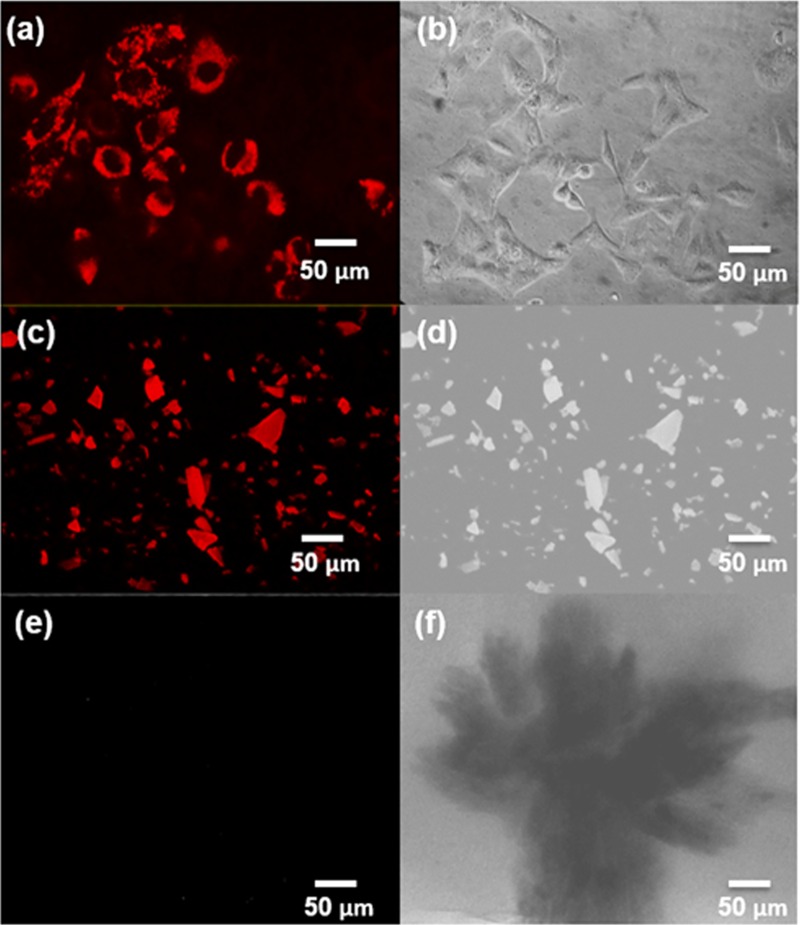
FIG 8.
(a) Fluorescence micrograph of HeLa cells stained with 30 μg/ml (0.13 mM) PbS2NPs. The nanoparticles exhibited red fluorescence, were evenly distributed in the cytoplasm, and did not stain the nuclei. (b) Phase contrast micrograph of HeLa cells treated with 30 μg/ml (0.13 mM) PbS2NPs. (c) Fluorescence micrograph of powdered PbS2NPs. (d) Phase contrast micrograph of powdered PbS2NPs. (e) Fluorescence micrograph of the lyophilized Idiomarina PR58-8. (f) Phase contrast micrograph of the lyophilized Idiomarina PR58-8.

