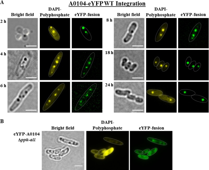
FIG 5.
(A) Expression of a genome-integrated A0104-eyfp fusion in R. eutropha. Cells were grown in NB medium at 30°C. Samples were taken at the time points indicated, were stained with DAPI for at least 10 min, and were imaged. The cell shapes are highlighted by thin white lines in the fluorescence images. The distribution of the polyP granules within the cell corresponded to that of wild-type cells. Note that the dark globular structures visible in bright field represent either PHB granules or polyP granules. (B) Localization of eYFP-A0104 in a polyP granule-free background of R. eutropha. Cells were grown in NB medium at 30°C. Expression of eYFP-A0104 in R. eutropha Δppk-all. The shapes of the cells are highlighted in fluorescence images by white dotted lines. The strong (yellow) fluorescence in one cell is not typical for the culture and is seen only in a minor fraction of cells. Bars, 2 μm.

