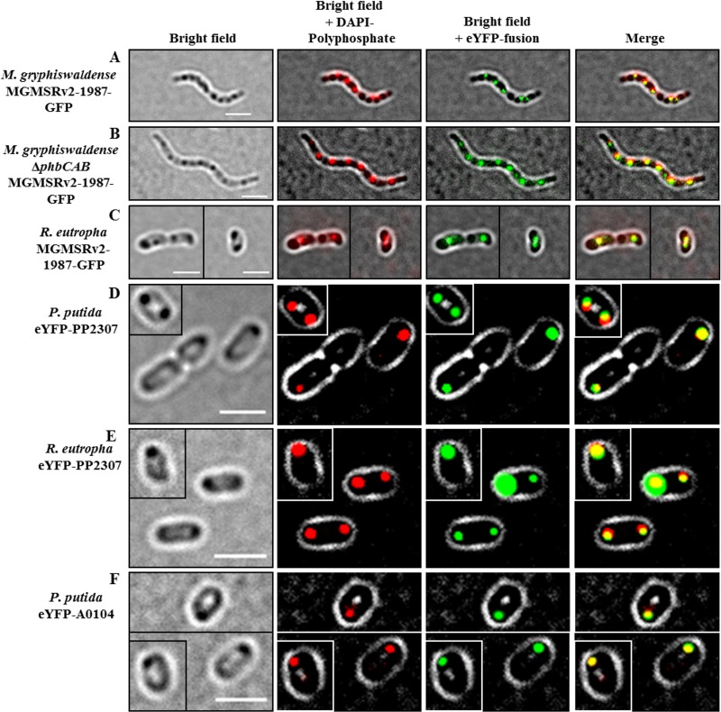
FIG 7.
Expression of CHAD domain-containing proteins MGMSRV2-1987-GFP, eYFP-PP2307, and eYFP-A0104 in different species and backgrounds. (A to C) Expression of MGMSRV2-1987-GFP in M. gryphiswaldense wild type (A), in a PHB-negative mutant M. gryphiswaldense ΔphaCAB background (B), and in R. eutropha (C). (D and E) Expression of eYFP-PP2307 in P. putida wild type (D) and in R. eutropha (E). (F) Expression of eYFP-A0104 in P. putida. Note the colocalization of the fusion protein with DAPI-stained polyP granules in all cases and no colocalization with the PHB granules that are visible in A and C as dark inclusions under bright field. Single cells in the frames of the images in C, D, E, and F correspond to cells of the very same culture and the same micrograph. To save space, cells were packed together in one image. Bars, 2 μm.

