Figure 5.
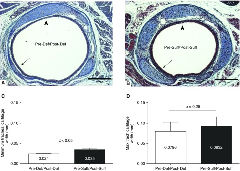
Effect of prenatal vitamin D deficiency on midtrachea cartilage morphology at 7 days. (A) Cross-section of the tracheal ring for a pup of a vitamin D–deficient dam (Pre-Def/Post-Def) shows segmental narrowing of the tracheal cartilage in the deficient mouse (arrow) when compared with (B) that of a pup from a sufficient dam (Pre-Suff/Post-Suff). The thickest part of trachea is comparable between the two tracheas (arrowhead; TriChrome stain [EMD Millipore, Billerica, MA], 100×). Scale bars, 250 μm. (C) Morphometric analysis showed thinner tracheal cartilage at its minimum dimension in the vitamin D–deficient (Pre-Def/Post-Def) group (P < 0.05). (D) Maximal cartilage thickness was similar between Vitamin D groups, emphasizing the heterogeneity of collagen deposition in the deficient mice. Results are expressed as mean (±SEM).

