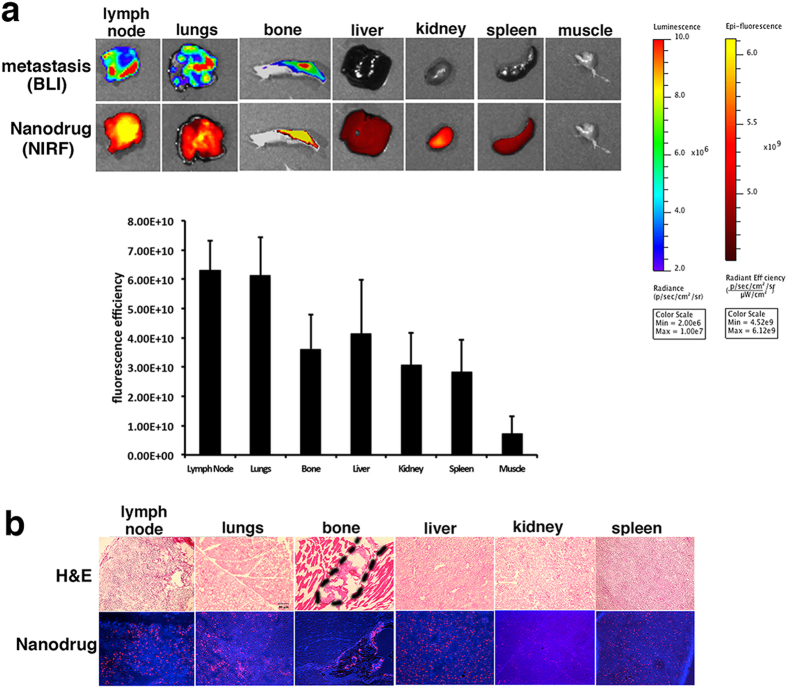
Figure 1. Nanodrug accumulation in metastatic organs.
(a) Top: Macroscopic imaging of metastatic burden (bioluminescence imaging, BLI) and nanodrug uptake (fluorescence reflectance imaging, Fl) in excised organs. Bottom: Quantitative analysis of fluorescence intensity in major organs). Metastatic organs were associated with high uptake of the nanodrug. Additional organs of high uptake reflected the natural pathway of metabolism and excretion of the nanoparticles. (b) Histology of tissue sections derived from excised organs. Top: H&E, bottom: fluorescence (Nanodrug: red, Cy5.5; nuclei: blue, DAPI). For lymph nodes and lungs, the whole tissue shown in the image is a metastatic lesion. The bone metastatic lesion is outlined in the image.

