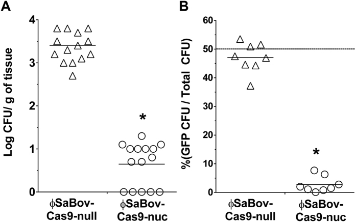
Figure 4. The efficacy of ϕSaBov-Cas9-nuc in in vivo murine skin infection.
The backs of C57BL/6 mice were intradermally infected with a suspension of (A) CTH96pGFP (1 × 10⁵ CFU) or (B) a mixture of CTH96pGFP and CTH96Δnuc (1:1, each at 5 × 10⁴ CFU) for 6 h, followed by treatment with the ϕSaBov-Cas9-nuc or ϕSaBov-Cas9-null at MOI of 500. After 24 h, infected skin was excised and homogenized. Viable cells were recovered by plating serially diluted homogenates onto BHI plate. The specificity of ϕSaBov-Cas9-nuc was evaluated by the proportion of viable cells expressing green fluorescent protein in total viable cells. Data points indicate the average of triple measurements in individual mice (n = 9). Asterisk indicates statistical significance in student t-test, compared to the results from ϕSaBov-Cas9-null (p < 0.001).

