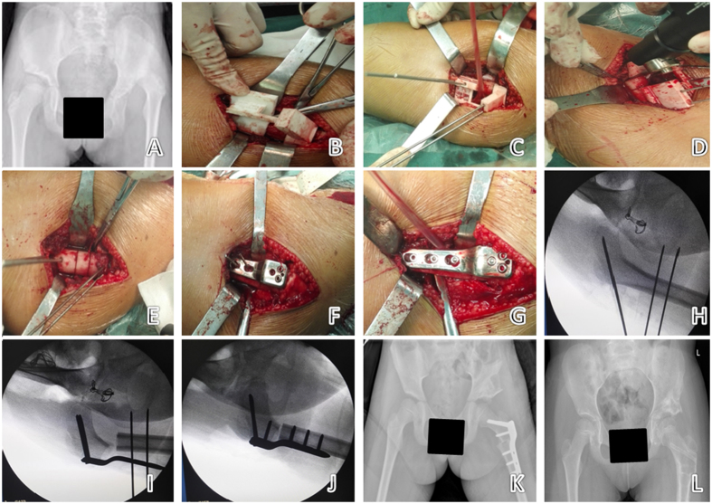
Figure 3. The navigation template applied in the operation on a 9-year-old girl with left DDH.
(A) Preoperative X-ray of the pelvis. (B–G) According to the simulation steps, the steps of the intraoperative process were completed as performed in the simulated operation. (H–J) Intraoperative use of C-arm X-ray to verify the direction of the needle and bone cutting form, consistent with preoperative planning (only 3 X-ray exposures). (K) X-ray of the pelvis one week after surgery. (L) X-ray of the pelvis 14 months after surgery.

