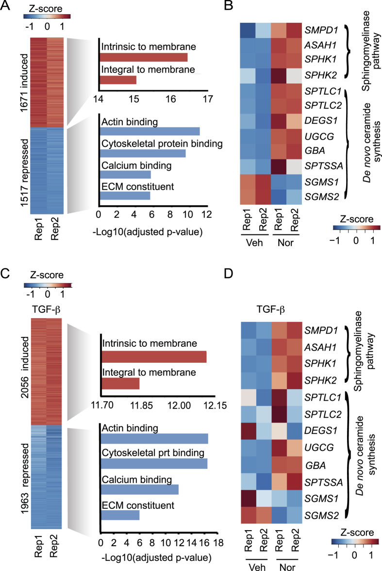
Figure 3. TCA treatment inhibits extracellular matrix constituents and regulates genes in the sphingomyelinase pathway.
(A) RNA-seq was performed on HSCs treated with ethanol vehicle or nortriptyline for 48 hours. Protein-coding genes induced (red) and repressed (blue) compared to vehicle are shown (at least 1.5 fold change in expression, false discovery rate < 0.0001). The relative expression (Z-score) of two replicates (Rep 1 and Rep 2) is shown. The most significant categories identified by GO analysis are shown for genes that were induced (red) or repressed (blue). (B) Expression of genes involved in the sphingomyelinase and de novo ceramide pathways following treatment with vehicle or nortriptyline. The relative expression (Z-score) is shown for two replicates (Rep 1 and Rep 2). (C) RNA-seq was performed on HSCs that were serum starved for 48 hours prior to treatment with TGF-β (2.5 ng/ml) and either ethanol vehicle or nortriptyline (27 μM) for 16 hours. Protein-coding genes that were induced (red) and repressed (blue) after nortriptyline treatment compared to vehicle are shown (at least 1.5 fold change in expression, false discovery rate < 0.0001). Relative expression (Z-score) for two replicates (Rep 1 and Rep 2) is shown. The most significant categories identified by GO analysis are shown for genes that were induced (red) or repressed (blue). (D) Expression of genes involved in the sphingolipid pathway following treatment with vehicle or nortriptyline in the presence of TGF-β. Relative expression (Z-score) is shown for two replicates (Rep 1 and Rep 2).

