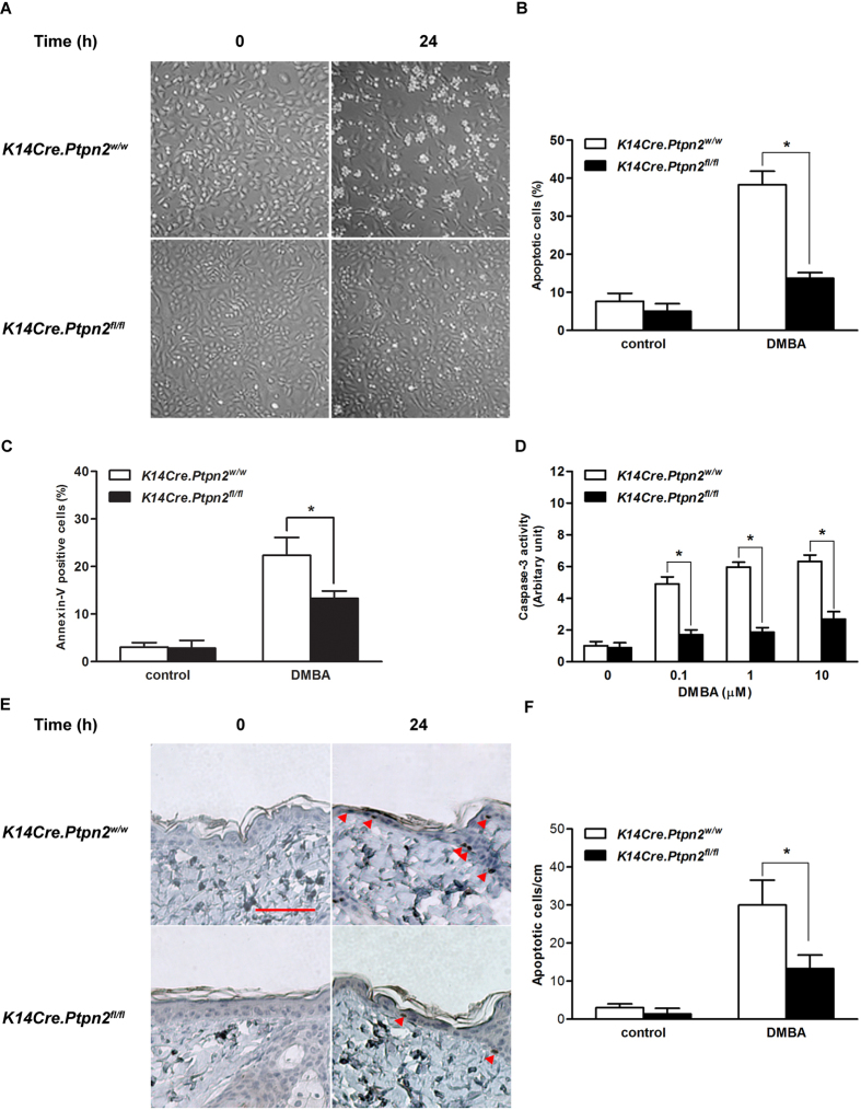
Figure 3. Effect of TC-PTP deletion on DMBA-induced apoptosis.
(A–C) Apoptotic response of primary keratinocytes obtained from the epidermis of K14Cre.Ptpn2w/w and K14Cre.Ptpn2fl/fl mice after 24 h of DMBA treatment. (A) Primary keratinocytes from both genotypes were cultured and treated with 100 nM of DMBA. (B) Quantitative analysis of percentage of apoptotic cells characterized by cell ballooning, nuclear condensation, and bleb formation. After 24 h of DMBA treatment, apoptotic keratinocytes were counted microscopically in at least three non-overlapping fields. Results are the mean ± s.d.m. from three independent experiments. *p < 0.05 by Mann-Whitney U test. (C) Flow cytometry analysis for apoptosis with Annexin V-FITC was measured after 24 h of 100 nM DMBA treatment. *p < 0.05 by Mann-Whitney U test. (D) Caspase-3 activity was measured after 24 h of DMBA treatment. *p < 0.05 by Mann-Whitney U test. (E–F) Apoptotic response of the epidermis from both genotypes. Groups of mice (n = 3) received a single topical application of DMBA (200 nmol) and sacrificed 24 h later. Skin sections were collected and apoptotic cells were quantified by immunostaining with caspase-3. (E) Representative staining of caspase-3 in the epidermis from both genotypes following treatment with DMBA. Scale bar: 100 μm. (F) Quantitative analysis of caspase-3-positive cells per centimeter of epidermis in both genotypes after DMBA treatment. *p < 0.05 by Mann-Whitney U test.

