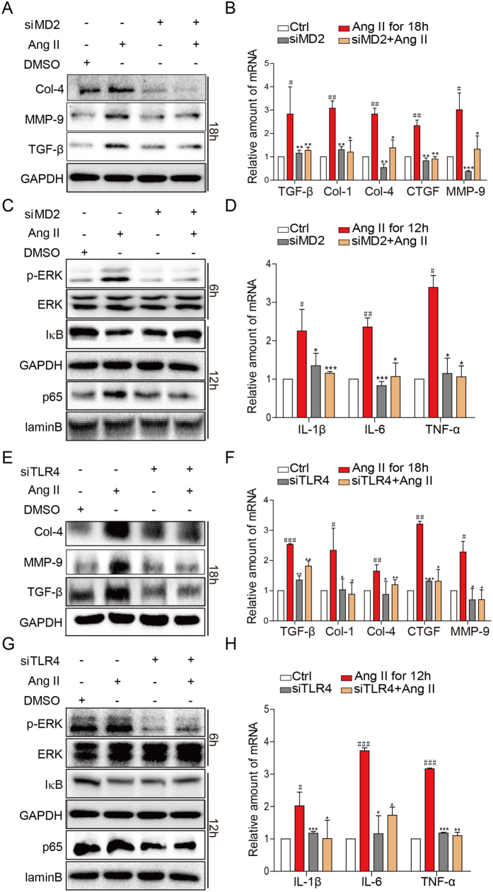
Figure 4. Ang II-induced inflammatory responses and signaling activation are dependent on the MD2 and TLR4.
NRK-52E cells were transfected with 1 μg MD2- or TLR4-specific siRNA in medium for 24 h, and stimulated with Ang II (1 mM) for different periods. (A,E) Representative Western blot analysis for marker proteins of fibrosis: Col-4 (collagen 4), MMP-9 (metalloproteinase 9), TGF-β (transforming growth factor β), GAPDH as loading control, n = 4 independent determinations. (B,F) The mRNA levels of TGF-β, Col-1 (collagen 1), Col-4, CTGF, MMP-9 were measured by real-time qPCR; values normalized to house-keeping gene β-actin. (C,G) Representative Western blot analysis for phosphorylated ERK (p-ERK), total ERK, and IκB from cell lysate; NF-κB p65 subunit analyzed from nuclear cell fraction; GAPDH and lamin B served as respective loading controls; n = 4 independent determinations. D,H) The mRNA levels of IL-1β, IL-6 and TNF-α were detected by real-time qPCR, values normalized to house-keeping gene β-actin. For data in (B,D,F, and H) values are reported as mean ± S.E.M. of n = 3; *P < 0.05, **P < 0.01, ***P < 0.001 versus Ang II-treated group; #P < 0.05, ##P < 0.01, ###P < 0.001 versus Ctrl. For panels A, C, E, and G, the gels were run under the same experimental conditions. Shown are cropped gels/blots (The gels/blots with indicated cropping lines are shown in Supplementary Figure S6).

