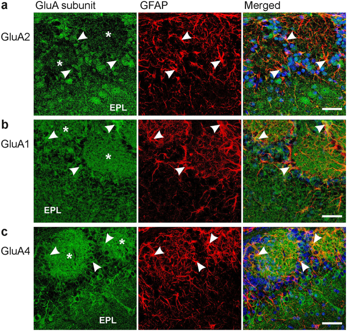
Figure 4. Immunostaining of AMPA receptor subunits in the olfactory bulb.
(a) GluA2 immunoreactivity (green) was detected in the external plexiform layer (EPL) and in cell bodies surrounding glomeruli. Glomeruli are indicated by asterisks. Moderate GluA2 immunoreactivity was also found in astrocytes highlighted by GFAP immunoreactivity (red), as indicated by yellow pixels in the merged image. Arrows point to astrocyte structures that were colabeled with GluA immunoreactivity. Nuclei were stained with Hoechst 33342 (blue). (b) GluA1 and GFAP colocalization. (c) GluA4 and GFAP colocalization. Scale bars: 20 μm.

