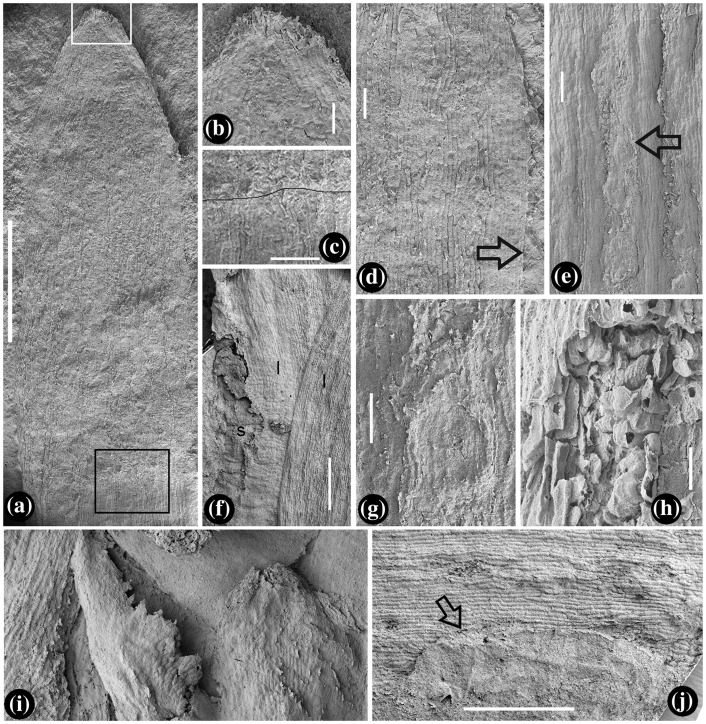
Figure 3.
Leaves and their details. SEM. (a) Abaxial view of leaf tip marked as 10 in Figure 2(a), showing the entire leaf margin and parallel veins. Bar = 1 mm, (b) leaf tip with papillae, enlarged from the white rectangle region in Figure 3(a). Bar = 0.1 mm, (c) leaf texture transitional from regular (below the line) to chaotic (above the line), enlarged from the black rectangle in Figure 3(a). Bar = 0.2 mm, (d) an adaxial view of a leaf, showing longitudinal epidermal cells and entire leaf margin (arrow). Bar = 0.1 mm, (e) an abaxial view of the leaf in Figure 2(d), showing well-defined alternating vein and intervein (stomata, arrow) zones. Bar = 0.2 mm, (f) leaf (l) clasping and diverging from the stem (s) with horizontal wrinkles. Note the leaf texture changes from the horizontal to longitudinal from the bottom up. Bar = 0.2 mm, (g) detailed view of the stomata arrowed in Figure 3(e). Bar = 0.1 mm, (h) a leaf with elongate epidermal cells (upper-left) and mesophyll aerenchyma. Bar = 50 μm, (i) a leaf in its earliest developmental stage, fringed with dentate protrusions. Bar = 0. 1 mm, and (j) leaf probably damaged by insect (arrow). Bar = 0.5 mm.

