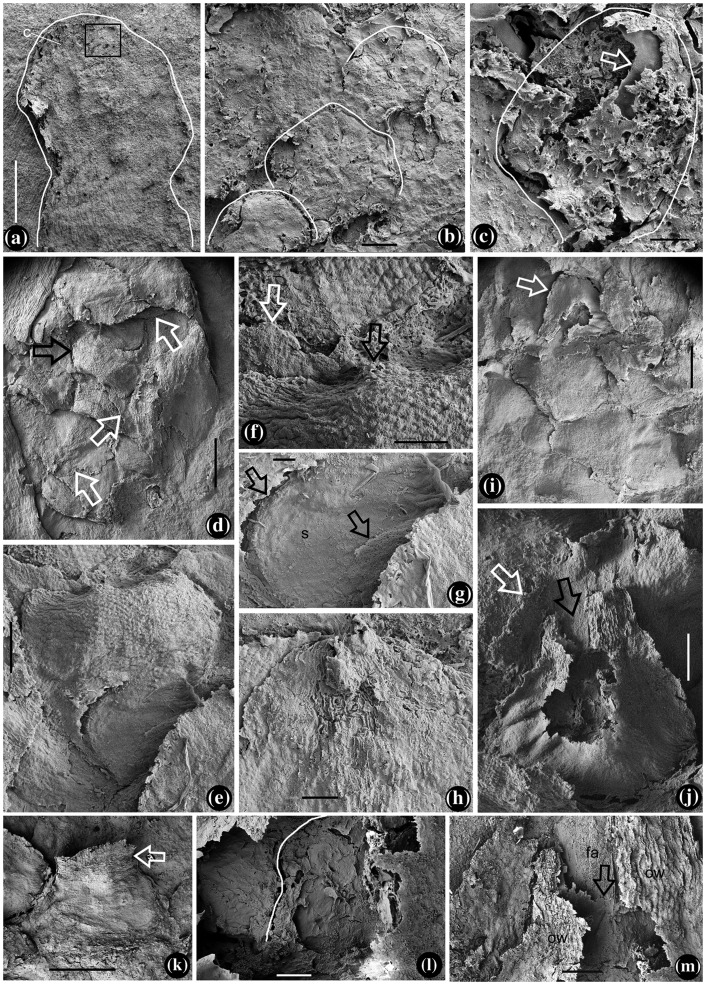
Figure 4.
Flower and aggregate fruits of Yuhania. SEM. (a) The immature flower in Figure 2(g), with a stout pedicel and spherical receptacle. Bar = 0.5 mm, (b) detailed view of the rectangle in Figure 4(a), showing outlines of the carpels helically arranged. Bar = 20 μm, (c) the sac-like carpel marked as C in Figure 4(a). Bar = 10 μm, (d) SEM view of the aggregate fruit in Figure 2(h), with helically arranged fruitlets. Bar = 0.5 mm, (e) one of the fruitlets from the aggregate fruit in Figure 4(d), with its seed exposed. Bar = 0.2 mm, (f) detailed view of the distal portion of the fruitlet in Figure 4(e). Note the cuspidate tip (black arrow), the greatest width near the distal of the fruitlet, and a bract (white arrow). Bar = 0.1 mm, (g) detailed view of the proximal part of the fruitlet in Figure 4(e), showing the broken fruitlet wall (arrows) and exposed seed (s) in the fruitlet. Bar = 50 μm, (h) rounded tip of a bract, note the longitudinal texture in the middle. Bar = 0.1 mm, (i) SEM view of the aggregate fruit marked as 2 in Figure 2(a) and shown in Figure S4(e) and (f). Bar = 0.5 mm, (j) a young ‘carpel’ with a broken tip (black arrow), wide base, and a bract in the background (white arrow). Note the empty space in the ‘carpel.’ Bar = 0.2 mm, (k) a young fruitlet with an extended terminus (arrow). Bar = 0.5 mm, (l) detailed view of young fruitlet shown in Figure 4(j), showing outline of a possible ovule (white line). Bar = 50 μm, and (m) detailed view of young fruitlet shown in Figure 4(j), showing ovarian wall (ow) fused (arrow) to the floral axis (fa). Bar = 0.1 mm.

