
Figure 1.
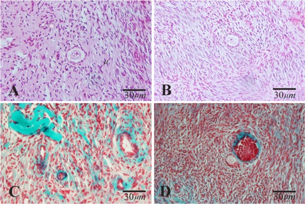
Light microscopic images of human ovarian cortical tissue after hematoxylin and eosin (A and B) and Masson Trichrome (C and D) staining before in vitro culture. A and C: non-vitrified group; B and D: vitrified group. The normal morphology of primary follicles was indicated in A, B and D