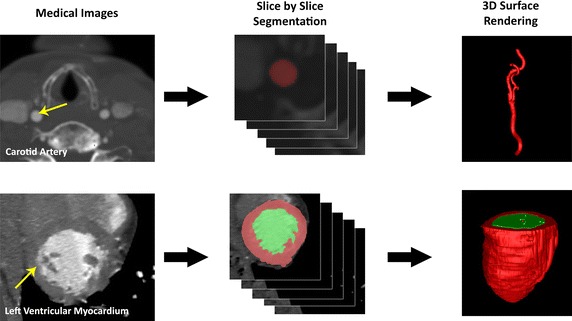
Fig. 2.

Medical image reconstruction of cardiovascular structures. Medical image reconstruction of cardiovascular structures. Computer tomographic angiography was carried out on the neck region of the patient whose carotid artery can be imaged at axial orientation for multiple slices. Segmentation based on the threshold of the blood vessel at various slices is performed in the initial stage. The segmented voxels can be grouped to form a three-dimensional anatomy and a mesh reconstruction based on the contours of these segmented regions is carried out (up). In a similar way, the left ventricle is imaged and ventricular chamber segmentation is performed. Then loft surface formation into a geometrical surface structure is enabled to give the anatomical model computationally (down)
