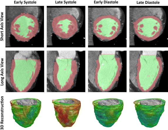
Fig. 3.

Geometrical reconstruction of left ventricle based on computer tomography. The images depict a short-axis (top) and long-axis (middle) scanning of the heart. The thickness of left ventricular endocardial and epicardial surfaces are traced with color mapping. Based on the myocardial segmentation, three-dimensional (3-D) reconstructions of the left ventricle (bottom) are prepare. The cardiacphases at the early, late diastole and systole are used as the time reference for hemodynamic assessment
