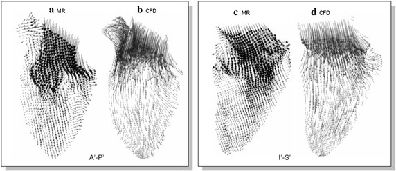
Fig. 6.

Flow fields of MR velocity imaging and CFD simulation. A 2D section of the velocity fields by the MRI modality and CFD simulation are displayed to characterize the flow within the left ventricle. The influxes of blood into the heart chamber as displayed by the two techniques generally possess the same kind of swirl nature. (Images from [36])
