
FIGURE 7.
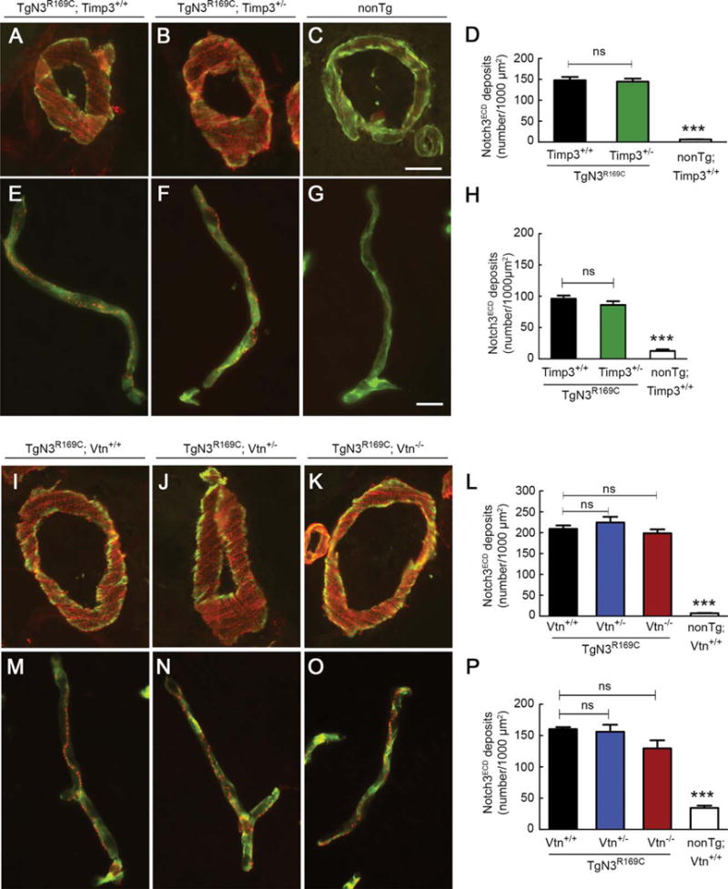
Notch3ECD deposition in brain vessels is unaffected by genetic reduction of Timp3 or vitronectin. Notch3ECD deposits were quantified in brain arteries and capillaries from mice with the indicated genotypes at 6 months of age (A–H) and 12 months of age (I–P). Representative images show brain arteries coimmunostained with anti-Notch3ECD antibody (red) and fluorescein isothiocyanate–conjugated anti–smooth muscle a-actin antibody (green; A–C, I–K) and brain capillaries coimmunostained with anti-Notch3ECD antibody (red) and antiperlecan antibody (green; E–G, M–0). Quantification of Notch3ECD deposit numbers per 1,000 μm2 in brain arteries (D, L) and brain capillaries (H, P) shows a significantly higher number of deposits in transgenic mice compared to nontransgenic (nonTg) mice, but a comparable number of deposits in TgN3R169C mice with normal or reduced expression of Timp3 (D, H) and normal, reduced, or no expression of vitronectin (L, P; n = 4–5 mice per genotype). Significance was determined by 1-way analysis of variance followed by Bonferroni post hoc test (***p < 0.001). Scale bars = 10 μm (A–C, I–K) and 15 μm (E–G, M–0). ns = nonsignificant.