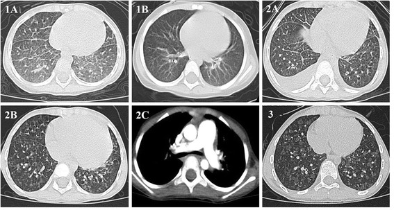
Fig. 1.

Lung CT showing the presence in both lungs of 1a diffuse ground-glass opacification predominant in lower region of lung and areas with smooth thickening of interlobular septum (on admission; Patient 1), 1b absence of abnormal pulmonary feature(after 1 month of treatment; Patient 1), 2a interlobular septal thickening and bilateral pleural effusion (1 year before admission; Patient 2), 2b diffuse poorly defined centrilobular nodules(5 days after treatment; Patient 2), 2c pulmonary artery (PA) with an enlarged diameter exceeding the aorta(5 days after treatment; Patient 2), and 3 diffuse poorly defined ground-glass centrilobular nodules(on admission; Patient 3)
