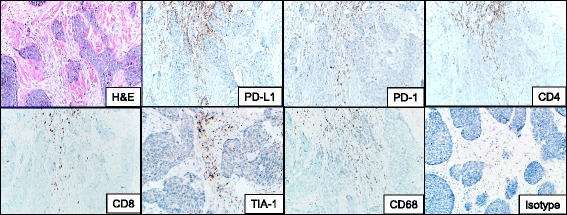
Fig. 3.

Immune elements in the microenvironment of a pre-treatment basal cell carcinoma from a patient who responded to anti-PD-1 therapy. The immune infiltrate abuts the tumor islands and is composed of a mixture of CD4 and CD8+ T-cells at a ratio of approximately 2:1. The CD8 cells are cytotoxic, as supported by the punctate cytoplasmic TIA-1 immunostaining. The lymphocytic infiltrate is accompanied by CD68+ macrophages. PD-1 is seen on approximately half of the lymphocytes present, and is immediately adjacent to PD-L1 expression in the tumor microenvironment, consistent with an immune microenvironment primed for potential response to PD-1/PD-L1 checkpoint blockade. PD-L1 is expressed predominantly on immune cells, rather than tumor cells in this example. H & E, hematoxylin and eosin, PD-(L)1, programmed death-(Ligand)1. 200× original magnification, all panels
