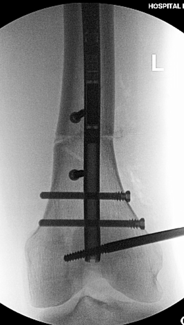
Figure 6.

The AP fluoroscopy shot shows the distal femur after successful distal interlocking with the varus deformity corrected. The peri-osteotomy blocking screws are positioned to prevent varus deviation during lengthening. The external half pin is also seen in the field.
