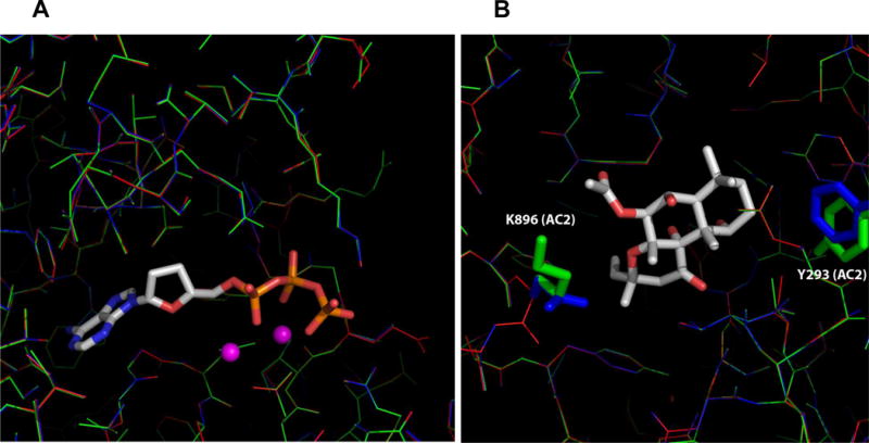
Fig. 5. Detailed-view of the ATP- and FS binding sites in mAC.

A, ATP substrate binding pocket. B, FS binding pocket. Two non-conserved residues, Tyr-293 and Lys-896, of AC2 and corresponding residues of AC1/AC5 within the 5 Å FS binding pocket are shown in stick mode. The color schemes for AC1, AC2, and AC5 model structure are blue, green, and red, respectively. ATP and FS are shown as stick models. Metal ions are shown as spheres.
